

Requirements for medical radiology and imaging centers in a complication epidemic
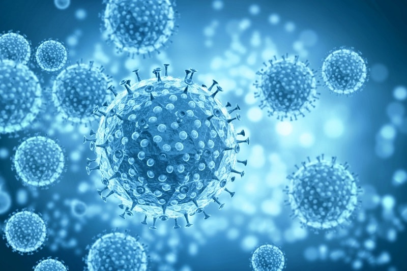
In December 2019, a new type of pneumonia caused by the unknown coronavirus (COVID-19) appeared in Wuhan, China. The virus spread rapidly around the world. Novel COVID-19 – infected pneumonia (NCIP) pneumonia is associated with fever, fatigue, dry cough, and shortness of breath.
Radiographic images of the chest of patients with Covid-19 are similar to those of other coronavirus syndromes, such as SARS and Mers. بررسی تغییرات تصاویر رادیولوژیکی بیماران مبتلا به سندرم حاد تنفسی یا سارس SARS (severe acute respiratory syndrome) و سندرم تنفسی خاورمیانه یا مرس MERS (Middle East Respiratory Syndrome) به محققان کمک میکند تا با مقایسه آنها با کرونا ویروس جدید کووید-19، به اطلاعات بیشتری دست یابند.
The aim of this study was to discuss and exchange information about the epidemiology and imaging findings of coronavirus syndromes. This information is based on NCIP imaging reports. In this study, the precautionary and safety measures of radiology department personnel against suspected patients with NCIP are reviewed.
History of SARS, Mers and Covid-19 coronaviruses
Coronaviruses are a large family of viruses that were first discovered in the 1960s. As of December 2019, six types of coronavirus were known to infect humans. Of these, four types of coronavirus caused mild respiratory symptoms, while Middle East respiratory syndrome (MERS) and acute acute respiratory syndrome (SARS) had acute respiratory symptoms.
In December 2019, a new type of coronavirus called COVID-19 was extracted from samples of the lower respiratory tract of several patients in Wuhan, China. These patients had severe pneumonia symptoms including fever, fatigue, dry cough, and respiratory distress.
Researchers believe that pneumonia caused by Covid-19 (NCIP) infection originated in the Wuhan Seafood Market. As of this writing, the coronavirus has spread to 28 countries. Evidence suggests that transmission of the corona virus can occur during the incubation period of the disease. The incubation period of a disease is the period of time when a person is unaware of their disease but is a carrier of the disease. The incubation period of the new Covid-19 virus is estimated at 5.2 days. This has led to the rapid spread of coronavirus. Also, after recovery from this disease, it is possible to transmit the disease through the saliva of a patient with NCIP for some time.
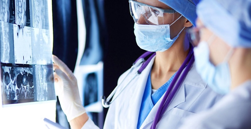
As of February 5, 2020, more than 25,000 cases of Covid-19 coronavirus have been reported, and the number of fatal patients is increasing. Medical imaging plays an important role in assessing the severity of Covid-19 infection. Radiologists should be well aware of the imaging findings of this new coronavirus.
Imaging features have already been identified in similar syndromes of this coronavirus. In this study, researchers investigate the epidemiological and radiological features of coronavirus syndromes based on known NCIP imaging features. Preventive and protective measures for radiology staff against suspected or coronavirus patients are also discussed.
Epidemiology and imaging findings of SARS coronavirus
The SARS coronavirus was first observed in 2003 in southern China. The virus quickly spread to 29 countries around the world. The SARS virus infected 8422 patients and killed about 11% of them. Coronavirus SARS was able to survive on dry surfaces for 24 hours, but lost its pathogenicity with a wide range of available disinfectants, such as Clorox and formaldehyde.
Early chest radiographs in people with SARS showed unilateral focal or multifocal opacities. Bubble turbidity was seen in the middle or lower peripheral areas of the lung with progressive dense (consolidation) masses over a period of 6 to 12 days in one or both lungs. CT scans of the lungs show ground-glass turbidity and consolidation in the affected areas of the lung.
Epidemiology and imaging findings of the coronavirus Mers
Coronavirus infection was first reported in 2012 in Jeddah, Saudi Arabia. Since then, about 2,500 people in 27 countries have been infected with the virus, with 30 percent dying. The risk of transmitting the disease to family members and health workers was low, but the epidemic of Hajj rituals was still widespread in Saudi Arabia.
Compared to human-to-human transmission in the SARS coronavirus epidemic, the transmission of the Merson coronavirus appears to have been of inhumane origin (bats and camels) and to have been transmitted from animal to human. In 83% of patients with MERS coronavirus infection, the initial radiograph showed some ground glass opacities.
CT scans of both lungs also showed glassy opacities that usually affected the lateral and lower parts of the lung. However, CT scans of patients with Mers coronavirus showed that consolidation and pleural effusion were uncommon and were seen in less than 30% of cases.
Coronavirus infections and clinical and radiological symptoms
Patients with COVID-19 infection have symptoms such as fever, cough, and shortness of breath. People usually feel tired but do not have symptoms such as runny nose, sore throat and diarrhea. A recent report in The Lancet examines coronavirus-19 pneumonia in 41 patients. Based on the imaging data in this study, lung abnormalities were observed bilaterally in 40 patients.
In the initial imaging reports of NCIP patients with severe symptoms, bilateral lung involvement with dense masses (consolidation) was observed. In patients in the intensive care unit, lung involvement with dense masses and ground-glass opacity was common.
Chest radiographs of a 61-year-old woman who died of coronavirus showed severe invasion of dense masses over a 7-day period. A separate report from 99 patients with coronavirus also found similar imaging findings. These findings showed bilateral lung abnormalities in 75% of patients and unilateral lung abnormalities in another 25%.
In another study of five members of a family infected with the coronavirus, bilateral glassy or grand glass facade was observed in the lungs. This involvement was more widespread in the lung parenchyma of older people. Imaging reports were very close to those of Sars and Mers.
Although no case of pleural effusion has been reported in coronavirus-19 pneumonia, pneumothorax has been reported in 1% of cases (one in 99 patients). In general, imaging findings in coronaviruses are highly nonspecific and may overlap with symptoms of H1N1 influenza, cytomegalovirus pneumonia, or specific pneumonia.
Severe clinical signs, a history of contact with a patient with Covid-19, or travel to Southeast Asian countries (such as China, South Korea, or Japan) are considered in the diagnosis of NCIP. Although more research is being done on the clinical and radiological aspects of COVID-19, imaging is considered an important component of patient management.
Requirements for radiology staff in dealing with patients suspected of having coronavirus
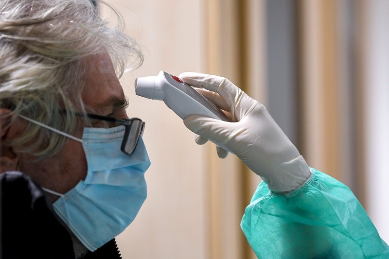
Radiology staff are at the forefront of dealing with patients suspected of having Covid-19. Imaging centers should have protocols in place to manage their staff against patients suspected of having Covid-19. The new coronavirus COVID-19 is highly contagious. Researchers believe the new coronavirus is transmitted through respiratory droplets. However, complete certainty about how the virus is transmitted from surfaces to humans is still under study. A thorough understanding of the virus transmission pathways is essential for the safety of patients and health care professionals.
Large droplets carry the risk of being transmitted up to 3 feet (91.44 cm) apart, but the virus may also be transmitted up to 6 feet (183 cm) away. For diagnostic imaging in people with NCIP, portable radiology equipment should be used to restrict patient transport.
Based on researchers’ experience with SARS, the use of a satellite radiography center and dedicated radiographic equipment can reduce the risk of transmitting individual infections. If it is necessary to transfer the patient to the radiology department, a surgical mask should be used during transportation.
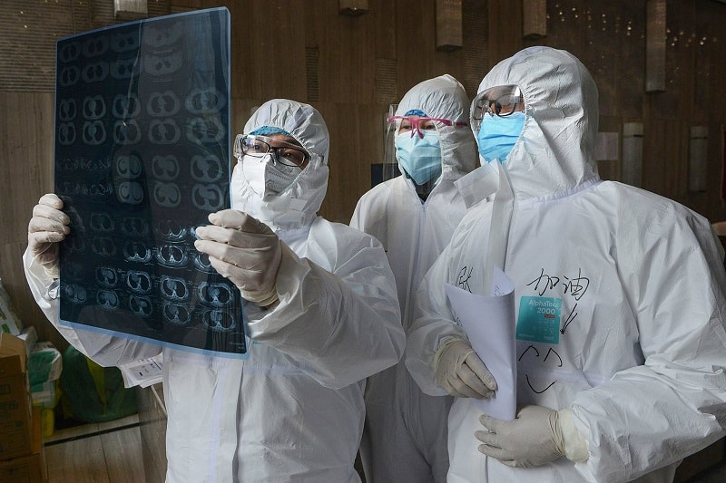
As of March 4, 2020, the World Health Organization has proposed the use of standard medical masks to protect the respiratory system. Other guidelines from the Centers for Disease Control and Prevention recommend the use of the N95 mask for people in direct contact with coronary heart disease.
In other instructions, it is recommended to use disposable, liquid-resistant insulating clothing (s) to protect personnel. In these instructions to avoid contact with respiratory droplets of patients with Covid-19, in addition to wearing guns; It is recommended to use a pair of disposable gloves on the clothes, cover the eyes with glasses and put a face shield on it.
In a study of 254 caregivers of patients with coronavirus SARS, safety and protective measures significantly reduced the incidence of the disease.
The tunnel (gantry) of CT and MRImachines, the probe of the ultrasound machine, the blood pressure bracelets, the keyboard, and the computer mouse for radiographic imaging should be disinfected after contact with each patient. According to the Spaulding classification at the Centers for Disease Control and Prevention and the Food and Drug Administration (FDA), these surfaces must either be washed with soap and water or with a low- or medium-grade disinfectant such as iodophor detergent. Disinfect with ethyl alcohol or isopropyl. Service personnel in imaging centers and institutions should be trained to properly disinfect surfaces.
0 دیدگاه