

Detected a very small portable ultrasound machine or a handhold?
Pleural effusion is a condition called pulmonary dehydration. The pleura is a thin membrane between the chest cavity and the lungs. In people with healthy lungs, there is a small amount of fluid between the lungs and the chest in the pleural membrane to prevent them from rubbing against each other because during respiration (tail), the volume of the lungs increases and they contract.
In fact, with excessive accumulation of fluid in the pleura, a person develops pleural effusion. This complication can be fatal and the patient should be treated immediately. Some people may develop pleural effusion due to underlying diseases such as pneumonia, heart failure, kidney disease, liver disease such as cirrhosis of the liver, and some cancers such as lung cancer and breast cancer.
This complication does not cause a specific symptom in all people and sometimes it can be seen only with imaging methods. This complication does not cause a specific symptom in all people and sometimes it can be seen only with imaging methods.
Diagnosis of pleural effusion is not easy and its symptoms may overlap with other lung diseases. In the first stage, the doctor listens to the patient’s lung sound using a stethoscope. After the patient’s clinical examination, the doctor usually requests a chest x-ray, which may be done by radiography, CT scan, or ultrasound.
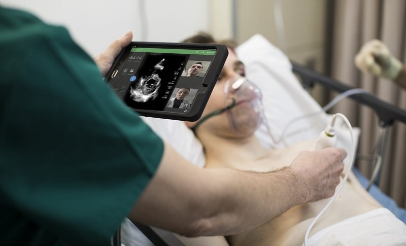
Sometimes the patient is in a bad mood and needs urgent medical attention. In this situation, the use of ultrasound for lung imaging is recommended because imaging with portable ultrasound devices is performed quickly and images are viewed simultaneously. Among portable ultrasounds, very small or handhelddevices have a very high capability for use in the emergency department and ICU. These devices are very light in weight and can be worn for a long time.
What are the diagnostic methods for pleural effusion?
CT scan is the gold standard for diagnosing pleural effusion. In addition to pleural evaluation, CT scans allow accurate evaluation of the chest wall, lung parenchyma, and mediastinum. One of the limitations of using the CT scan method is the lack of this device in any area. This device is very expensive and patients have to wait for a CT scan, which delays the diagnosis. This places a huge constraint on critically ill and accidental patients.
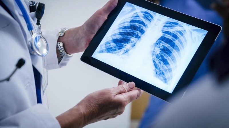
The primary method for examining pleural effusion is plain chest radiography. For pleural effusion to be detected on chest radiograph, at least 175 ml of fluid must have accumulated in the pleural space. Lateral radiographs can also detect this complication with 75 ml of accumulating fluid in the pleural space.
However, not all patients can have a radiograph. This is especially important in patients admitted to the intensive care unit (ICU) and accidental patients in the emergency department. On the other hand, some technical issues affect the quality of radiographic images. These problems are especially noticeable when the patient is lying in bed. These problems include chest movement during imaging, patient rotation, and short film-to-body distances.
Comparison of ultrasound and radiography in the diagnosis of pleural effusion
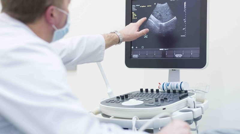
Ultrasound has been used to diagnose pleural effusion since the late 1960s. A 1976 study used A-mode to detect pleural effusion with 93% sensitivity. Other studies over the years have shown that ultrasound has performed better than radiography in diagnosing pleural effusion. However, the use of ultrasound is not as common as chest radiography.
Compared to ultrasound, radiography has the advantage of evaluating the skeleton as well as the lung and mediastinum parenchyma. In addition, radiography can examine tubes and chest catheters. Ultrasound, on the other hand, has the ability to reveal opaque lesions such as effusions, atelectasis, and dense masses. For years, the ability of ultrasound to detect effusions and use them in imaging procedures has been limited. This has increased the success of imaging procedures such as thoracosynthesis, drainage, and biopsy, and reduced mortality.
In recent years, studies have shown that in measuring the volume of the effusion, ultrasound produces better results than radiography. Among radiologists, ultrasound is known as a sensitive and specific method for diagnosing pleural effusion. Ultrasound is a fast, low-cost method that the portable device is portable and can accurately detect pleural effusion without the use of ionizing radiation. The use of ultrasound may be expanded as the first choice for evaluating patients suspected of having pleural effusion if further studies are reported on the accuracy of the diagnosis of pleural effusion.
Advantages and limitations of Handheld portable ultrasound in the diagnosis of pleural effusion
In the emergency department, the time of examination and diagnosis is very important. The parameters of pleural effusion evaluation are identified by ultrasound techniques in a few minutes. Therefore, portable ultrasounds can be used to assess pleural effusion in emergencies.
Ultra-portable or handheld ultrasound is a portable and lightweight ultrasound device that has a lower price than other portable devices. This device, like other portable ultrasound devices, has the ability to detect fluids in the pleural space and complications such as pleural effusion with appropriate accuracy.
These devices weigh about 300 to 600 grams and usually have strong batteries. The doctor can easily move the handheld devices with her for a long time In patients with subcutaneous emphysema and obese patients, the diagnosis of pleural effusion by ultrasound is limited. Also, in patients whose chest is covered with a wound, bandage, or catheter, a complete ultrasound may not be performed. Another important point is the issue of educating physicians about ultrasound diagnoses. Learning pleural effusion detection techniques is not very complicated and can be taught in a short period of time.
Due to the many advantages of handheld portable ultrasound devices over their limitations, it is recommended that these devices be used for early detection of free fluid in the body in the emergency department.
3 comments